Impact
The Fabrica created a stir in the anatomical world. Some, especially the Parisian anatomists who had been at the forefront of the Galenic revival, were extremely critical of Vesalius’ work, and accused Vesalius of misunderstanding Galen. Others, including Gabriele Fallopius at the University of Padua, enthusiastically adopted Vesalius’ approach and were soon pointing out instances in the Fabrica where Vesalius himself had relied on classical authority. The illustrations themselves also made quite an impression, and were promptly copied and inserted into other anatomists’ texts, much to Vesalius’ disgruntlement.
Charles V was pleased with Vesalius’ accomplishment, and promptly made him one of his court physicians. Vesalius returned briefly to Italy, where he carried out a series of public dissections before entering imperial service. He then followed Charles throughout the Emperor’s travels in northern Europe, serving as a physician and surgeon. After Charles’ abdication in 1556, Vesalius entered the service of his successor, Philip II. He moved to Spain with the imperial court and remained there until 1564, when he departed on a pilgrimage to Jerusalem for reasons that have never been completely understood. He fell ill on the return voyage and died on the Greek island of Zakynthos, supposedly near the town of Zante.
Vesalius was not the first anatomist to challenge a reliance on textual authority and to assert that there were errors in Galen’s anatomical descriptions. By the time the Fabrica was published, other anatomists, most notably Berengario, were stressing the importance of first-hand observation over what the ancient authorities had written. The Fabrica was, however, a truly remarkable achievement. Vesalius’ integration of text and image, his emphasis on the hands-on dissection of human bodies and willingness to challenge the established medical canon, and the sheer beauty of the completed Fabrica ensure its enduring significance as a work of both anatomical and artistic significance.
Realdo Colombo

Realdo Colombo (c.1510-1559) was born in the northern Italian city of Cremona, and received a liberal arts education in Milan before spending seven years as an apprentice to the Venetian surgeon Giovanni Antonio Lonigo. He left Lonigo’s service to begin studying medicine at the University of Padua, where he was one of Vesalius’ students. Their relationship was good enough that in the 1543 edition of the Fabrica Vesalius referred to Colombo as his friend, and Colombo served as interim Professor of Anatomy and Surgery during Vesalius’ sojourn in Basel.
When Vesalius returned, the relationship became sour. In his anatomy lessons at Padua, Colombo had taken it upon himself to point out the anatomical errors in Vesalius’ new work. Vesalius did not take kindly to this. In his 1546 “China Root Epistle” Vesalius stated that Colombo had been “incompletely educated,” and all mentions of Colombo were excised in the second edition of the Fabrica.
In 1545 Colombo accepted a teaching position at the University of Pisa at the invitation of Cosimo de’Medici, but was only there for three years before moving on to the Papal Medical School in Rome. During his time there he met the artist Michelangelo, and the two of them made plans to produce a new illustrated anatomical atlas that would be comparable to the Fabrica. This unfortunately never came to pass, as Michelangelo died before the project could be completed. The only illustration in Colombo’s De re anatomica, published posthumously in 1559, is the title page. It is not as elaborate as that of the Fabrica, but uses the same imagery of the anatomist performing a dissection with his own hands.
Despite Vesalius’ assertions to the contrary, Colombo was a skilled anatomist. He made significant contributions toward the understanding of pulmonary circulation, and also described the relative positions of the kidneys correctly.
Juan Valverde
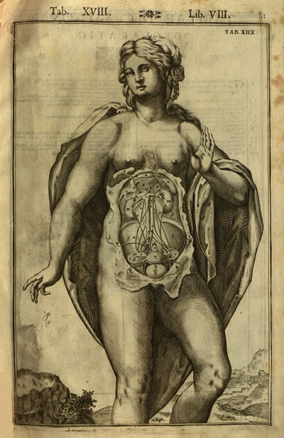
Juan Valverde de Amusco’s Historia de la Composicion del Cuerpo Humano, which was first published in 1556 in Rome, is one of the most notable plagiarisms of Vesalius. The majority of the copperplate engravings are copies of the Fabrica’s illustrations with the backgrounds excised. Although Vesalius himself said that the text appeared to be written by someone who had little practical experience with dissection, Valverde’s text proved to be quite popular and was published in multiple editions.

Valverde’s text did include some original illustrations, which were probably drawn by Gaspar Becerra and engraved by Nicolas Beatrici. The one shown here refers to Saint Bartholomew, who is sometimes said to have been flayed alive, and in Renaissance art is often depicted holding his own skin.

This title page is from an edition printed in Rome in 1560, and contains all of the conventional images that we would expect to find in a 16th century anatomical publication. The three images at the bottom all relate to aspects of anatomical instruction – on the left a professor is providing instruction on the bones, the center shows a formal anatomical lesson complete with consultation of an authoritative text, and the rightmost image shows a smaller group dissecting a female corpse, probably referring to one of the private anatomies that were used to supplement large public displays. The pig and the monkey perched on the top of the cartouche allude to the fact that those animals were frequently used in anatomical instruction.

This title page from the 1586 Venetian edition shares many features with the 1560 version, including a depiction of a pig and a monkey, and scenes of dissection. The skin of a satyr that stretches over the top of the central cartouche is the most striking difference. This refers to the Greek legend of Apollo and Marsyas. Marsyas was a satyr who engaged in a music contest with Apollo, with the condition that the winner could inflict whatever punishment they desired upon the loser. When Marsyas lost, Apollo skinned him alive.
Casserius
Julius Casserius (1561-1616) was another one of the exceptional anatomists who taught at the University of Padua. He trained under Vesalius’ successor Fabricius ab Aquapendente, and in 1604 became the University’s professor of anatomy. He commissioned beautiful copperplate illustrations to accompany his anatomical texts, but most of them were unpublished when he died in 1616. The German physician Daniel Bucretius acquired the plates from Casserius’ heirs with the intention of using them to illustrate Adrian van der Spieghel’s De humani corporis fabrica, and published them under the title Julii Casserii Placentini Tabulae anatomicae LXXIIX in 1627.
The initial seventy-eight plates acquired by Bucretius were drawn and engraved by Joseph Maurer. He then commissioned an additional twenty, which were drawn by the Venetian artist Odoardo Fialetti and engraved by Francesco Valesio. These illustrations, which are among the most beautiful anatomical images ever created, show the influence of the Vesalian woodcuts. Like Vesalius’ musclemen, the anatomical figures in Casserius’ plates are depicted in lifelike positions against naturalistic landscapes, and are posed in a manner that is reminiscent of Italian paintings and statuary.
Ambroise Paré

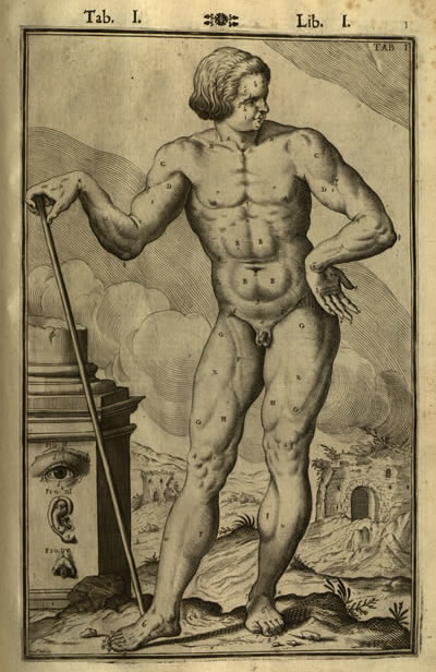
Illustrations that were clearly modeled after the original Vesalian woodblocks continued to appear in medical texts for the next two hundred years. These images come from a seventeenth century English edition of the collected works of Ambroise Paré. Paré was born in 1510 and was a contemporary of Vesalius, although he never received a doctorate in medicine. He entered military service and worked as a barber surgeon, and later went on to serve in the courts of Henry II, Charles IX, and Henry III. He is most well remembered for his method of treating gunshot wounds, which he developed during his time on the battlefield. When he ran out of boiling oil to cauterize wounds with during the Siege of Turin, Paré used an improvised dressing that consisted of egg yolk, oil of roses, and turpentine. The men he treated with this method healed much better than those whose wounds were cauterized, and from then on Paré endorsed the new method.

Paré’s writings proved very popular, and went through multiple editions in several languages. The edition shown here is a 1678 English edition that was printed by a woman, Mary Clark. The anatomical illustrations are clearly small copies of Vesalius’ originals.